


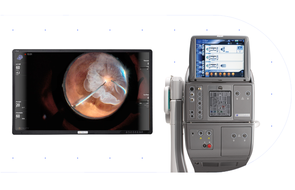
Redefine what’s possible with NGENUITY®
The True Digital 3D Visualization System


It’s time to go beyond the limits of an analog microscope and experience the superiority1,2* of a truly digital visualization system so you can:
Visualize
your surgery with increased confidence due to enhanced detail, magnified and digitally optimized to help achieve the best possible results2
Integrate
your Alcon Vision Suite with DATAFUSION to access your most important information the moment you need it
Streamline
OR workflow and transform collaboration with staff through a heads-up surgical view
Improve
the way you operate with greater ergonomics, heads-up viewing, and optimized patient outcomes with integrated Digital Image Guidance by Alcon®3-8
Experience
Alcon’s best-in-industry customer service, training, and support every step of the way9,10
* Compared to analog microscopes
Superior visualization of NGENUITY®
Now with Digital Detection
Digital Detection allows you to see beyond the capabilities of your analog microscope.
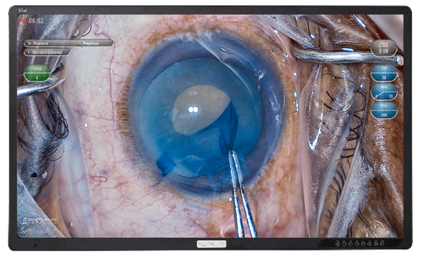
Tissue Detail Mode* (TDM)
Visualize detail and depth like never before during the most delicate steps of your procedure
Capsule Clarity Mode* (CCM)
Edge detection for greater ability to identify capsule construction, compared to standard microscopes, during your toughest cases11†
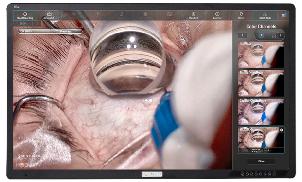
MIGS Mode*
Benefit from up to 34% greater contrast, allowing for better identification of TM, leading to enhanced precision and efficiency with MIGS placement12**
Digital Detection allows you to see beyond the capabilities of your analog microscope.
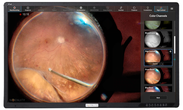
Tissue Detail Mode* (TDM)
Visualize depth and detail like never before during vitreous and membrane removal
Performance Green Mode* (PFG)
Specific digital mode for green dyes allowing for greater visualization and contrast of membranes during macular work
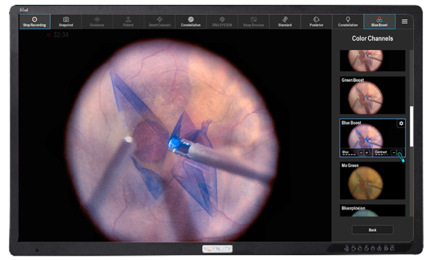
Blue Boost Mode* (BBM)
Digital image feature for blue dyes allowing for greater visualization and contrast during macular work
See like never before...
View with expanded clarity
Up to 5x extended depth of field delivers a crisp focus across an expanded surgical space and a deeper range of details.2,*,†
Enhanced precision and accuracy
minimizes the need to manually refocus during the procedure, promoting efficiency and reducing operating time.13,14
Immerse in a magnified view
Up to 48% increased magnification provides an expansive view, improving the ability to perform intricate surgical tasks.2,*,†
Accentuates details at high magnification beyond the capabilities of an analog microscope for delicate procedures.
Distinguish every detail
Up to 42% increased depth resolution enables your ability to resolve fine details when managing challenging pathologies.2,*,†
Enhances your ability to distinguish various layers of the anterior anatomy for refined tissue manipulation.2
...with a true digital experience
NGENUITY® provides enhanced visualization with MIGS Mode for the treatment of glaucoma
With the enhanced visualization benefits of NGENUITY®, discover how MIGS Mode can benefit from up to 34% greater local contrast, allowing for greater precision and efficiency in identifying the trabecular meshwork (TM) during MIGS placement.12*
MIGS Mode Off
MIGS Mode ON

With DATAFUSION, NGENUITY® integrates all analog microscopes and your existing Alcon equipment to perform cataract procedures with precision, efficiency and a reduced risk* of misalignment errors for your patients.2,3,5,6

*Image Guided integration eliminates manual marking, reducing the risk of misalignment errors.
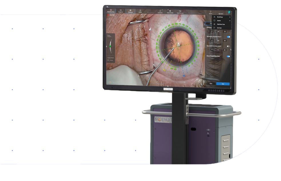
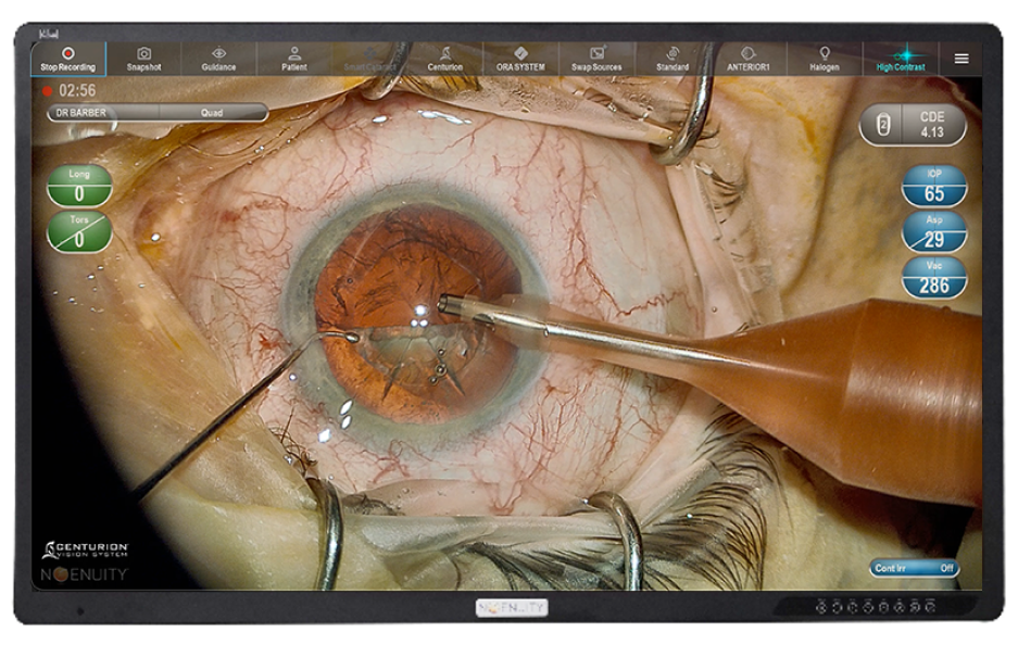
Alcon Digital Image Guidance
Drive Efficiency and Reduce Manual Error – Enable surgeons and surgical teams to access key surgical data on the SMART Solutions platform through a centralized surgical view in the OR.
NGENUITY®:
- Provides integration with ARGOS®, connecting your clinic to the OR
- Introduces Alcon Digital Image Guidance, helping surgeons achieve optimal lens placement for increased precision and alignment6-8,16,17
- No calibration required and hands-free navigation of image guidance steps17
- Faster overlay tracking for greater efficiency1
- Increased reticle resolution for greater precision17
- Stereo reticle display for dominant eye independence1
- Reduces OR complexity, eliminating additional equipment and cable
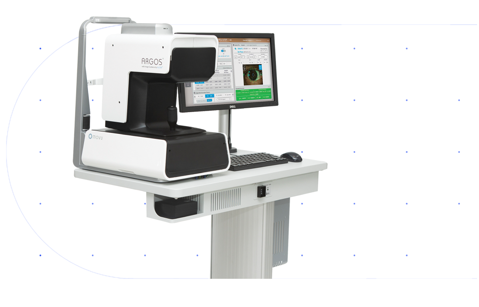
ARGOS® Biometer
Connecting Clinic to the OR – ARGOS® is integrated with the Alcon Vision Suite to ensure better outcomes with greater efficiency.6-8,16
ARGOS®:
• Provides integration with ARGOS®, connecting your clinic to the OR
• Streamlines your pre-op and post-op workflow by eliminating the need for time-consuming manual data entry
• Removes potential for transcription errors
• Executes your IOL plans with image guided precision through the NGENUITY® system
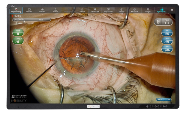
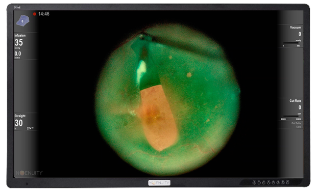
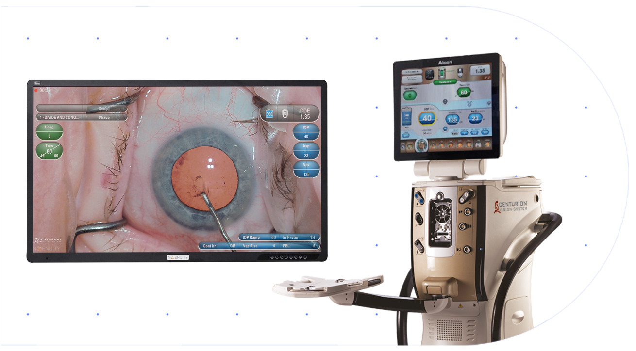
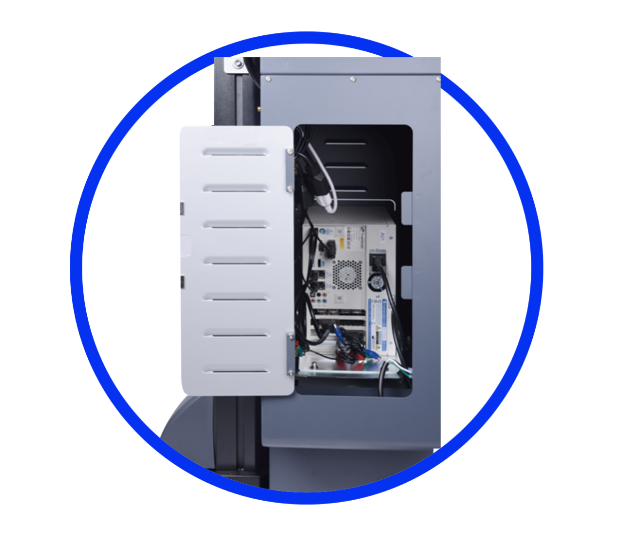
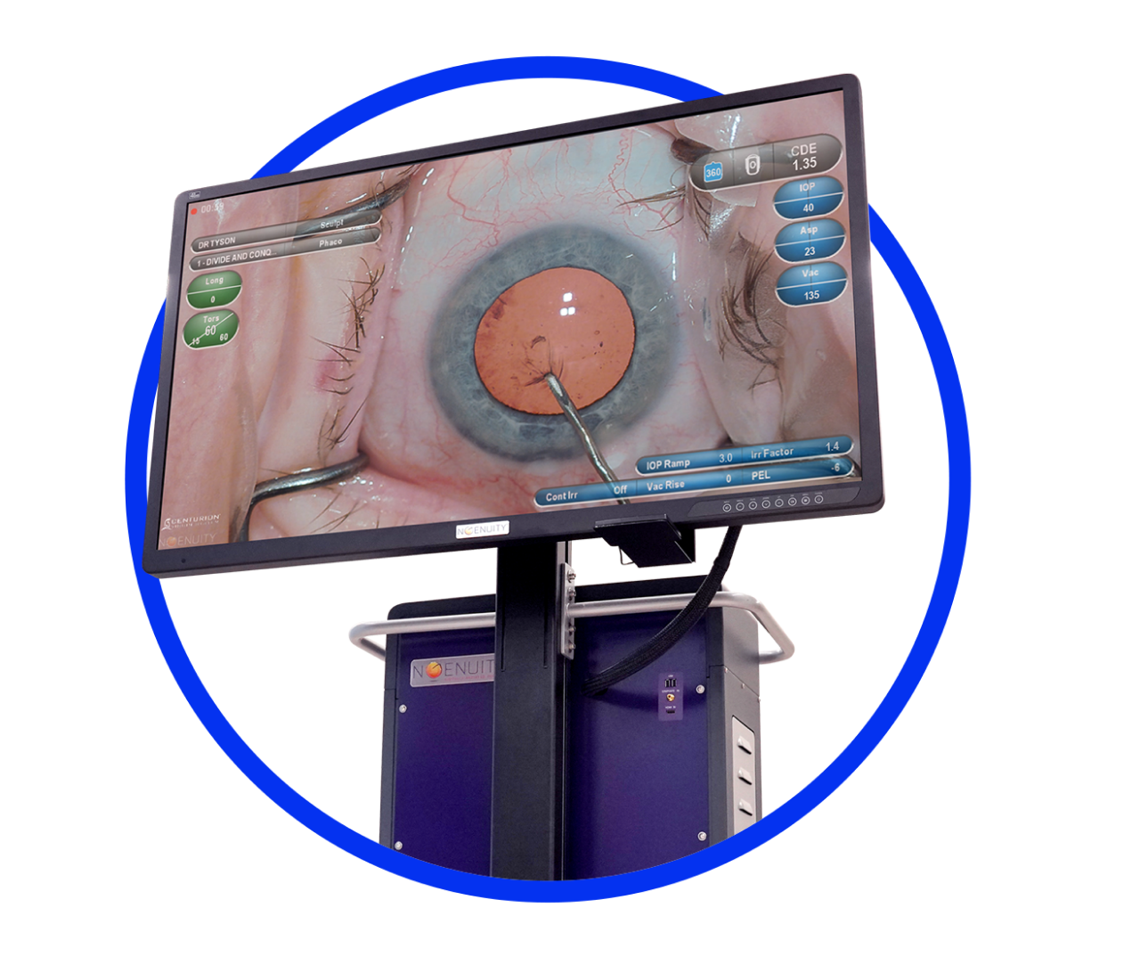
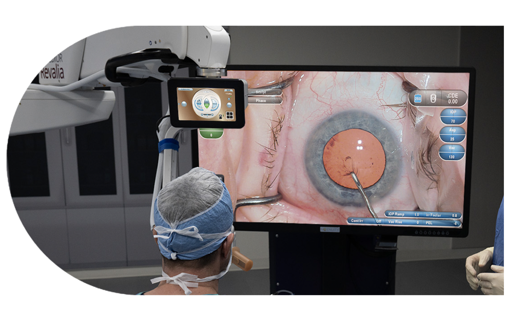
CENTURION® DATAFUSION
A Centralized View, Tailored To You – CENTURION® DATAFUSION delivers a real-time view of surgical parameters and system performance providing more information during critical surgical steps, allowing you to keep your eyes on what's most important.18
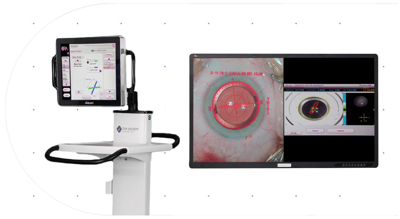
ORA VerifEye+TM DATAFUSION
Seamless Access To Diagnostic Data – Expand your intraoperative diagnostic usage and decision-making with ORA® with VerifEye+™ cart and aberrometer data displayed directly on the NGENUITY®.
Experience better reticle representation on the live camera image, compared to oculars, for an improved patient image.
CONSTELLATION® DATAFUSION
A Centralized View, Tailored to You – CONSTELLATION® DATAFUSION delivers a real-time view of surgical parameters and system performance providing more information during critical surgical steps, allowing you to keep your eyes on what's most important.8
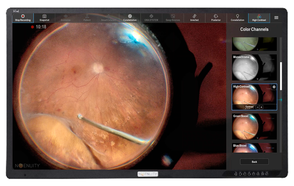
Color profiles
Personalized digital profiles provide more flexibility to customize your surgical approach with a multitude of applications.1*

Light temperature profiles
Easy-to-use thumbnails allow you to customize the light profile during the surgical procedure, no matter what microscope you’re using.1

Less microscope light
This matters. Digital image processing offers the ability to operate under low lighting conditions for increased patient comfort and a reduced risk of phototoxicity.5,15
Custom image profile
Link your CONSTELLATION® procedural steps with your preferred image modes for greater efficiency and enhanced visualization at every step.2,13,14
*Compared to analog microscopes
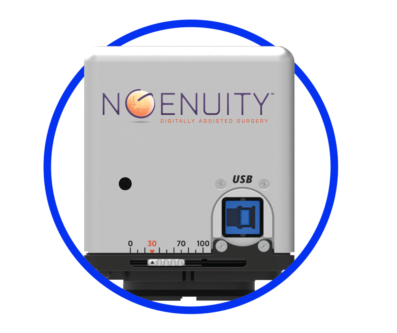
Versatile HDR surgical camera1
See every detail with high dynamic range imaging that merges underexposed images with overexposed images for greater detail recognition and contrast.
Set your own depth of field for image optimization with the upgraded, adjustable aperture.
Designed attachment flexibility that adapts to almost all ophthalmic surgical microscopes so you can experience the benefits of digital visualization while maximizing your investment.
Features:
• True stereoscopic 3D imaging
• Two full HD Sensors (1920x1080)
• 60 fps per eye
High-speed image processor1
Delivers real-time image processing for unparalleled surgical viewing.
Provides personalized color and light temperature adjustments through easily accessible image thumbnails.
Enables 3D/2D recordings for impactful podium presentations and teaching situations.
1.5 Enhancement Modes with Digital Detection:
• Capsule Clarity Mode (CCM)
• MIGS Mode
• Tissue Detail Mode (TDM)
• Available for both Anterior Segment and Posterior Segment
• Blue Boost Mode (BBM)
• Performance Green Mode (PFG)
• Digital Image Guidance
Features:
• Stream 3D/2D to multiple displays simultaneously
• Real-time video recording and playback
• Picture in picture or split-screen processing
Enhanced OLED surgical display1
Compared to LCD display, OLED provides more immersive 3D viewing with better contrast, higher brightness and a wider color range.
Compared to LCD display, OLED provides more immersive 3D viewing with better contrast, higher brightness and a wider color range.
Features:
• 4K OLED resolution (3840×2160p)
• 55-inch screen size
• 60 fps for each eye
Increase comfort with the potential for reduced risk
Surgeon Risk: Improved Ergonomics and Reduced Musculoskeletal Discomfort
NGENUITY® can help resolve ergonomic issues associated with conventional microscopes. Surgeons are 5x more likely to report reduced discomfort/pain, improved posture, and improved overall comfort.3,4,19-21*
Patient Risk Factor: Less Microscope Light
Experience up to 4x lower illumination levels without compromising visual outcomes during surgery.22
In a study of hybrid vitrectomy cases, NGENUITY® enabled a reduction of light from the microscope, chandelier light, and endo-illuminator by 40%, 60% and 20%, respectively.5
Reduced Endo-Illumination
With NGENUITY®, surgeons were able to potentially decrease the risk of patient phototoxicity by reducing endo-illumination, while still working comfortably using 23-, 25- and 27-gauge instruments.4

Less Dye Needed
With NGENUITY®, surgeons could perform chromovitrectomies using lower concentration of dyes, or no dye at all, which may reduce potential toxic side effects of these products.11,23
*In a multivariable model of questionnaire responses from 64 surgeons indicating the odds of reporting an improvement in pain since introducing the HUD in the operating room for those who used HUD in >50% of their cases (mean years of HUD use=2.3, P=0.029).
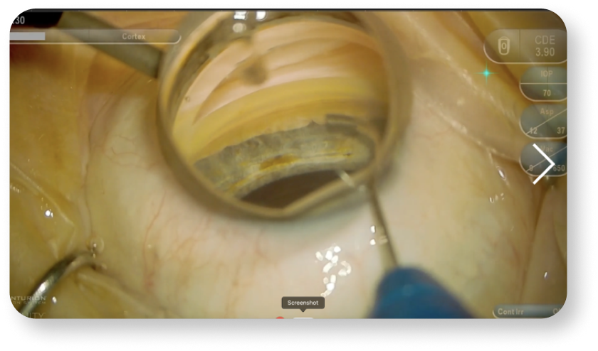
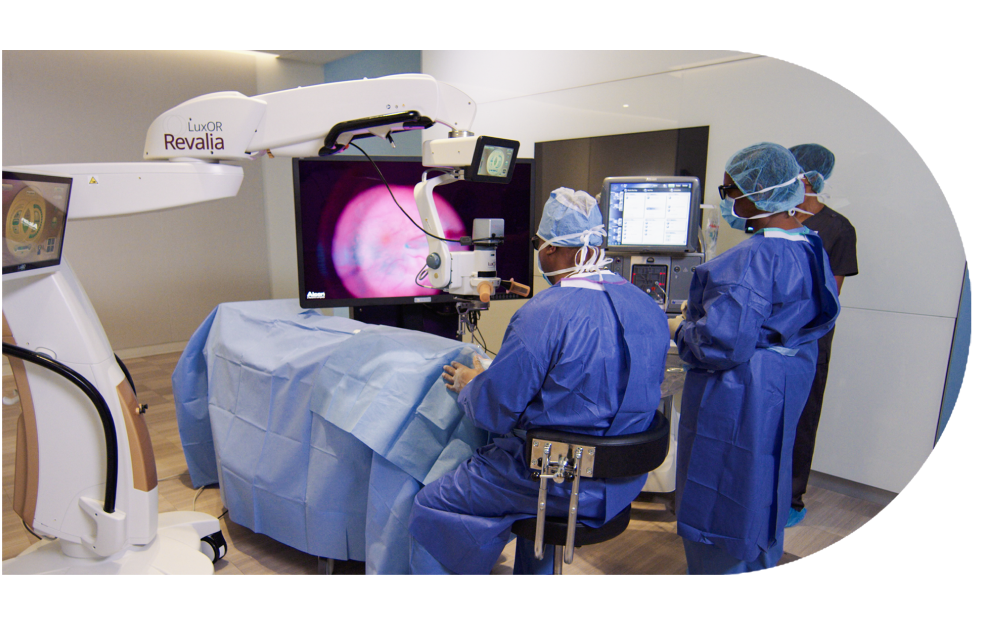
Streamline OR workflow
Software enhancements and improvements to the system help to simplify your OR workflow13, allowing staff and residents to see with the same depth, focus and clarity as the surgeon.11
Digital image enhancement
Experience the difference of digital with an increase in magnification, depth of field and image resolution for crisp detail.2,*,†
* Compared to analog microscopes including the Leica Proveo 8 and Zeiss OPMI LUMERA 700 scopes.
† Specified performance was achieved at maximum system magnification with an aperture setting of 30% open and viewing distance of 1.2 meters.
Integrated image guidance
Connectivity with DATAFUSION to your complete surgical suite delivers an on-screen, real-time view of surgical parameters and system performance for greater information sharing during critical surgical steps.
Powers collaboration and teaching
Accelerates knowledge transfer through a shared view, improving the collective learning experience in the OR while immersing the whole surgical team in a comprehensive view of the procedure, allowing them to anticipate your next steps.11
Freedom from oculars
The benefit of heads-up surgery improves ergonomics, compared to traditional analog binoculars, while digitally assisted visualization takes your experience to the next level.4
Advanced recording system
2D and 3D recording allow for instant surgical case review, with the option to save and share your surgical case recordings outside the OR, giving audiences a true representation of your experience.1
Benefit from Alcon’s industry-leading customer service, training, and support every step of the way9,10
With Alcon, surgeons and staff have access to resources to make the transition to NGENUITY® seamless.
Alcon Experience Academy
For relevant training content from industry thought leaders
IMPORTANT PRODUCT INFORMATION
Caution: Federal (USA) law restricts this device to sale by, or on the order of, a physician.
Indication: The NGENUITY® 3D Visualization System consists of a 3D stereoscopic, high-definition digital video camera and workstation to provide magnified stereoscopic images of objects during micro-surgery. It acts as an adjunct to the surgical microscope during surgery displaying real-time images or images from recordings.
Warnings: The system is not suitable for use in the presence of flammable anesthetics mixture with air or oxygen. There are no known contraindications for use of this device.
Precautions: Do not touch any system component and the patient at the same time during a procedure to prevent electric shock. When operating in 3D, to ensure optimal image quality, use only approved passive-polarized glasses. Use of polarized prescription glasses will cause the 3D effect to be distorted. In case of emergency, keep the microscope oculars and mounting accessories in the cart top drawer. If there are any concerns regarding the continued safe use of the NGENUITY® 3D Visualization System, consider returning to using the microscope oculars.
ATTENTION: Refer to the user manual for a complete list of appropriate uses, warnings and precautions.
References
1. NGENUITY® 3D Visualization System User Manual.
2. Alcon Data on File, 2017.
3. Zhang Z, Wang L, Wei Y, Fang D, Fan S, Zhang S. The Preliminary Experiences with Three-Dimensional Heads-Up Display Viewing System for Vitreoretinal Surgery under Various Status. Curr Eye Res. 2019 Jan;44(1):102-109. doi: 10.1080/02713683.2018.1526305.
4. Eckardt C, Paulo EB. Heads up surgery for vitreoretinal procedures: An experimental and clinical study. Retina. 2016;36:137-147.
5. Kita M, Mori Y, Hama S. Hybrid wide-angle viewing-endoscopic vitrectomy using a 3D visualization system. Clin Ophthalmol. 2018;12:313-317.
6. Shammas HJ, Ortiz S, Shammas MC, et al. Biometry measurements using a new large-coherence-length swept-source optical coherence tomographer. J Cataract Refract Surg. 2016;42:50-61.
7. Whang W, Yoo Y, Kang M, et al. Predictive accuracy of partial coherence interferometry and swept-source optical coherence tomography for intraocular lens power calculation. Sci Rep. 2018;8(1):13732.
8. Shammas HJ, Shammas MC, Jivrajka RV, Cooke DL, Potvin R. Effects on IOL power calculation and expected clinical outcomes of axial length measurements based on multiple vs single refractive indices. Clin Ophthalmol. 2020;14:1511-1519.
9. Alcon Data on File, 2022 [US surgical strategic insights]
10. Market Scope: 2021 Annual Sponsored US Cataract Surgeon Survey Report. May 2021.
11. Moura-Coelho N, Nascimento J, Henriques J, Medeiros MD. Three-dimensional display systems in ophthalmic surgery – a review. European Ophthalmic Review. 2019;13(1):31-36.
12. Alcon Data on File, 2022.
13. Berquet F, Henry A, Barbe C, et al. Comparing heads-up versus binocular microscope visualization systems in anterior and posterior segment surgeries: a retrospective study. Ophthalmologica. 2020;243(5):347-354.
14. González-Saldivar G, Chow DR. Optimizing visual performance with digitally assisted vitreoretinal surgery. Ophthalmic Surg Lasers Imaging Retina. 2020;51(4):S15-S21.
15. Hamasaki I, Shibata K, Shimizu T, et al. Lights-out surgery for strabismus using a heads-up 3D vision system. Acta Med Okayama. 2019;73(3):229-233.
16. Tamaoki A, Kojima T, Hasegawa A, et al. Clinical evaluation of a new swept-source optical coherence biometer that uses individual refractive indices to measure axial length in cataract patients. Ophthalmic Res. 2019;19:1-13.
17. ARGOS® Biometer User Manual.
18. VERION™ Digital Marker M User Manual.
19. Cheng TC, Yahya MFN, Mohd Naffi AA, Othman O, Seng Fai T, Yong MH, Wan Abdul Halim WH, Mustapha M, Che Hamzah J, Md Din N, Bastion MC. Evaluation of Three-Dimensional Heads up Ophthalmic Surgery Demonstration From the Perspective of Surgeons and Postgraduate Trainees. J Craniofac Surg. 2021 Mar 24. doi: 10.1097/SCS.0000000000007645.
20. Palácios RM, de Carvalho ACM, Maia M, Caiado RR, Camilo DAG, Farah ME. An experimental and clinical study on the initial experiences of Brazilian vitreoretinal surgeons with heads-up surgery. Graefes Arch Clin Exp Ophthalmol. 2019 Mar;257(3):473-483. doi: 10.1007/s00417-019-04246-w.
21. Weinstock RJ, Ainslie-Garcia MH, Ferko NC, Qadeer RA, Morris LP, Cheng H, Ehlers JP. Comparative Assessment of Ergonomic Experience with Heads-Up Display and Conventional Surgical Microscope in the Operating Room. Clin Ophthalmol. 2021 Jan 29;15:347-356. doi: 10.2147/OPTH.S292152.
22. Rosenberg ED, Nuzbrokh Y, Sippel KC. Efficacy of 3D digital visualization in minimizing coaxial illumination and phototoxic potential in cataract surgery: Pilot study. J Cataract Refract Surg. 2021;47:291-296.
23. Alcon Data on File, 2020.